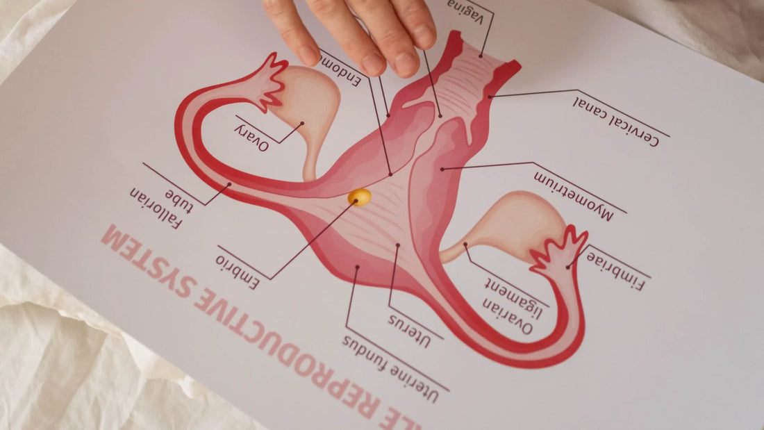
What is the anatomy of a uterus?
The uterus is located between the urinary bladder anteriorly and the rectum posteriorly. The average dimensions of the uterus in an adult are 8 cm long, 5 cm across, and 4 mm thick. The uterine cavity has an average volume of 80 mL to 200 mL. The uterus subdivides into three segments, namely: the body, the cervix, and the fundus.
The uterus has three tissue layers which include the following:
- Endometrium: the inner lining and consists of the functional (superficial) and basal endometrium. The functional layer responds to reproductive hormones. When this layer sheds, this results in menstrual bleeding. If there is damage to the basal endometrium, this can result in the formation of adhesions and fibrosis (Asherman syndrome).
- Myometrium: the muscle layer and is composed of smooth muscle cells.
- Serosa/Perimetrium: the thin outer layer composed of epithelial cells.
At the upper corners of the uterus, the fallopian tubes (tiny passageways for the eggs to travel) connect the uterus to the ovaries (oval-shaped organs in the upper right and left of the uterus where the eggs are kept). They produce, store, and release eggs into the fallopian tubes in the process called ovulation. Once the egg is in the fallopian tube, tiny hairs in the tube's lining help push it down the narrow passageway toward the uterus. The ovaries (OH-vuh-reez) are also part of the endocrine system because they produce female sex hormones such as estrogen (ESS-truh-jun) and progesterone (pro-JESS-tuh-rone).


